

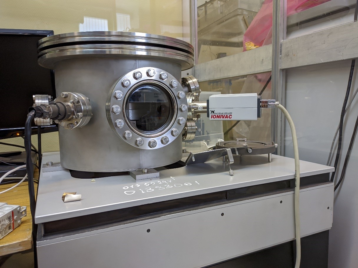
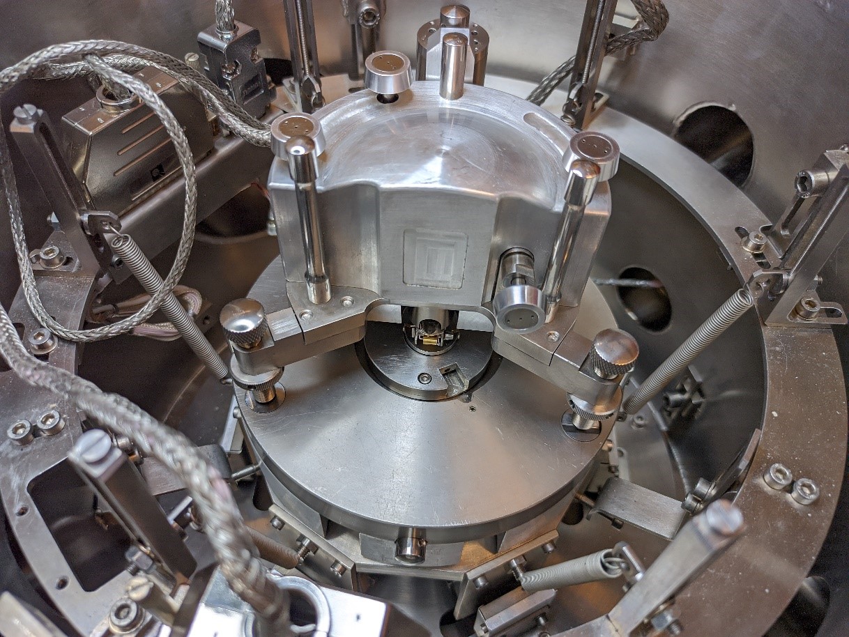
Allows you to investigate the morphology of the sample by atomic force and tunneling microscopy. The scanning head of the microscope is located in a vacuum chamber, which provides research in medium and high vacuum conditions. The presence of active and passive vibration protection allows achieving high measurement accuracy. The presence of a specialized measuring head makes it possible to measure the morphology of objects in a liquid medium. Allows to implement the following measurement methods:
Atomic force microscopy (contact, non-contact and semi-contact modes): study of the surface morphology of the sample.
Magnetic force microscopy: study of the distribution of the magnetic moment over the surface of the sample.
Kelvin probe method: measuring the distribution of the surface potential (contact potential difference) over the surface of the sample.
Piezoresponse force microscopy: The PFM technique is based on the inverse piezoelectric effect, which is a linear relationship between the electric field and mechanical deformation.
Scanning Tunnel Microscopy: Study the morphology of conductive samples. Allows you to get an image with atomic resolution. It also allows one to obtain tunneling current-voltage characteristics of the samples.
Examples of images obtained during the study:
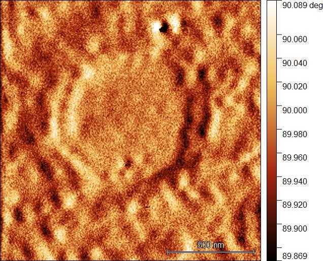
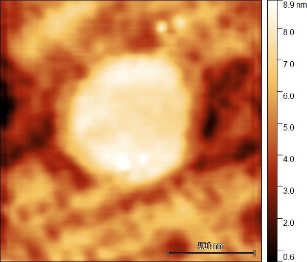
Images of nickel arachinate nanoclusters (image size 1×1 μm), formed under Langmuir monolayers of arachidic acid, obtained in the semi-contact mode of displaying the surface topology and phase contrast